Report on “Modern Equine Dentistry” with Alice Hughes
Firstly, we must thank Jen Morris for organising another fantastic zoom session, this time with Alice Hughes BVMS BAEDT MRCVS.
Jen started off by introducing Alice, who Jen used to teach at Pony Club and now Alice is married with children and is her vet. Alice is part of a large practice in Aberdeen called Ardene House Vet Practice Ltd and has lots of exciting, new techniques.
Alice started off by informing us she is not a “specialist” vet in Equine Dentistry, but she has a special interest in it and is spending a lot of her evenings studying for other qualifications in this area. She is a member of British Association of Equine Dental Technicians (BAEDT) and is studying to become an advanced member. There are Equine Dental Specialists who travel around running clinics and Alice works closely with them for a couple of days when they are up every few months and she will also travel down to Dorset to gain further experience.
ANATOMY:
 Alice started by explaining that the horse’s cheek teeth (molars) come further back than most realise, and the point of the facial crest is where the fourth cheek tooth lays so there are several more teeth behind. We saw a skull where the bone had been removed to show how much space the teeth occupy of the horse’s head, the image was of an older horse, Alice said in a younger horse, the teeth are close to the orbit!
The teeth are numbered the same way as humans with the mouth being split into quarters. You split the teeth from the central incisors and from the upper jaw and the mandible, for instance,
Left side uppers: central incisor = 101, lateral incisor = 102, corner incisor = 103. The canine = 104 (found in male horses but sometimes females), we can see small wolf teeth on the image which are numbered 105, and the cheek teeth (three premolars and three molars) are 106/7/8/9/110/111.
The right side would be 201, 202 and so on, the mandible would be numbered 301 onwards for the left and 401 onwards for the right.
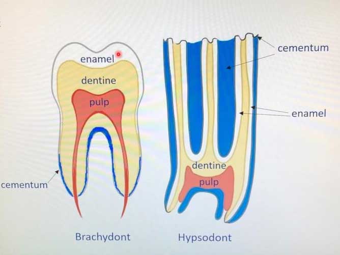
We then looked at the tooth anatomy, as you can see from the picture I have added, Alice compares a human tooth to an equine:
Humans are Brachydont and equines are Hypsodont’s. As we know, once we have our adult teeth, they stay whereas a horse’s teeth keep erupting throughout its life. The image shows a young horse’s tooth, the older it gets, the less tooth there will be.
Our teeth are white as they have the enamel covering them, whereas you can see all three dental tissues of a horse’s tooth on the chewing surface. The cement (infundibula) is on the sides and in the middle with white ridges of enamel which are responsible for grinding hay as they chew and are also responsible for the sharp points as the tooth erupts. Then between you have the dentine. The dentine covers the pulp and the pulp horns. For the mandible cheek teeth, they are more rectangle in shape compared to the upper jaw and have no channels of cement.
Wolf teeth should never be removed for under 2.5 years of age that is when the first cheek tooth sheds, so it often sheds the wolf tooth with it. They are commonly picked up when the horse is going to be backed as they get their teeth checked so the horse is ready to go. If they are small, Alice is tempted to leave them in unless they are causing problems. There is no evidence/written papers to say they cause a problem. Alice will remove them if they are further forwards than the cheekteeth or if there’s not enough room to fit a double bridle.
AGEING:
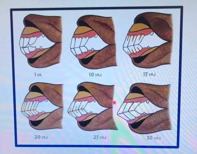 As everything must be passported, it takes a lot of the ambiguity out of ageing but there are a lot of ways to age a horse from looking. The longer the tooth, the older the horse. A younger horse is easier to age as a horse will lose their baby centrals at 2.5 years, laterals at 3.5 and corners at 4.5 years. The Galvayne’s groove is a staining of food that comes in at 10 years at the top of the corner incisor and is halfway through at 15 years, but it is not accurate. A better way is to look at the incisors infundibula (cup), if the cup is not in wear in the corners, the horse is in its fifth year. The infundibula leave the teeth at 6 for the centrals, 7 for the laterals and 8 for the corners. In a foal, the corner incisors and the fourth cheek tooth come in at 9 months of age. A horse is born with three cheek teeth which are then lost at 2.5, 3 and 4 years of age.
ROUTINE DENTISTRY:
Alice recommends routine dentistry every six months for horses under 8 and over 18. Younger horse’s teeth are constantly erupting up to 8mm per year for a 5yo! When the horse is in its teens, this slows to about 1mm per year.
When to get them checked: quidding, nasal discharge, weight loss, smell, ridden issues, bitting problems, swellings, and anxiety around the head.
A dental exam is more about looking with mirrors than rasping. The upper jaw is wider than lower teeth so outside of the upper jaw and inside of bottom need filing as there’s nothing to stop these enamel points getting longer and sharp!
Make sure dentists are qualified – this is not enforceable for people to be qualified to do dentistry so check! The two companies are, The British Association of Equine Dental Technicians (BAEDT) who work with BEVA and have a list of “EDT” on their website and The Worldwide Association of Equine Dentists (WWAED) who also have a list of qualified and registered dental technicians.
When examining, most people now use power rasps. They need a good speculum/gag, need a head torch, rest if horse sedated. The mouth needs to be flushed out so you can see clearly how the tooth is. With using a mirror, a clean mouth, and a steady head, you can see things like fillings or decay.
COMMON PROBLEMS:
Wolf teeth – if there is a gap between them and the first cheek tooth, these would cause problems when ridden so they are best to be removed.
Strong/you take a strong contact, the first cheek tooth will show wear so if there are wolf teeth present, these should come out. When extracting teeth, the horse must be sedated, and local anaesthetic given.
Ulcers – a result of sharp enamel points where the horse needs to more regular dental checks. The inside of the bottom jaw can cause ulceration to tongue and the outside upper can ulcerate the fleshy cheek.
 Overgrowth – Alice explained that they cannot just cut the tooth down to the gumline due to the pulp horn and if you do, the tooth will bleed and could even die. They must file down gradually, letting the sensitive pulp horn repair. This is seen as the black bit, which is the secondary dentine but as soon as the black bit goes, the next colour is blood! If this happens, it is agony for the horse and fillings would be needed and the tooth would probably die over the next couple of years.
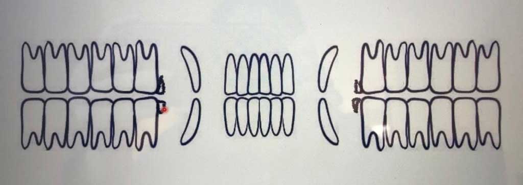
Diastemata & Periodontal Disease – Diastema is the food getting trapped and the Periodontal is the painful gum disease. There are two types of diastema, a “valve” diastema, and an “open” diastema.
A valve diastema is more commonly found in younger horses where the teeth meet at the top but then there is a gap lower down and the food gets in, but then cannot get out again and as you can see, the gums have started to recede from gum disease.
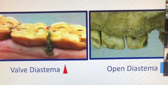 An open diastema as seen in the picture with the parallel gaps from biting surface to gumline (more on the left of that picture – the big gap is where a tooth has been removed). Here, food can get out and you do not tend to get the severe gum disease with these. An open diastema is more commonly found in the older horse.
Treatment for a valve diastema would be to clean it out and then widen the gap which changes the shape of a “V” to a rectangle. This in turn lets food out. They used to cut the rectangle to the gumline but now they just cut a groove and the idea is, if the food is fully cleared out, with the wider groove, the food should just pass over.
Peripheral caries (decay) – this is a release acid which erodes the cement around the outside of the teeth and is often found with diastema’s as bacteria feed off the food that is trapped, which releases the acid. However, as teeth are constantly erupting if you can change the ph. in the mouth, healthy cement with grow.
Infundibula Caries – (only on the top teeth). When teeth are forming, some cement will/can be missing which is most common in the 9’s (oldest teeth in the mouth) or the first cheek. To treat, they would drill out all the rotten food and cement, clean, etch the surface so the filling will then stick and fill. If left untreated, decay would spread to the pulp horns and the tooth can fracture. The specialist equine dentists have been using this technique for about 10 years now and there are studies out now proving its effectiveness.
Pulp Exposure – black lines which is the dentine that overlaps the pulp horns. In a cheek tooth, a horse can have five, six or seven pulp horns which communicate with each other in different ways i.e. one and five will often communicate. If too much pulp is exposed, over time the food will collect in the gaps and will decay which will mean a tooth extraction is necessary. The horse would be sedated with nerve blocks given so they cannot feel anything. The vets then spread the gap at the front and back, grab the tooth and wiggle it gradually and slowly down.
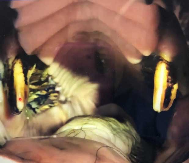
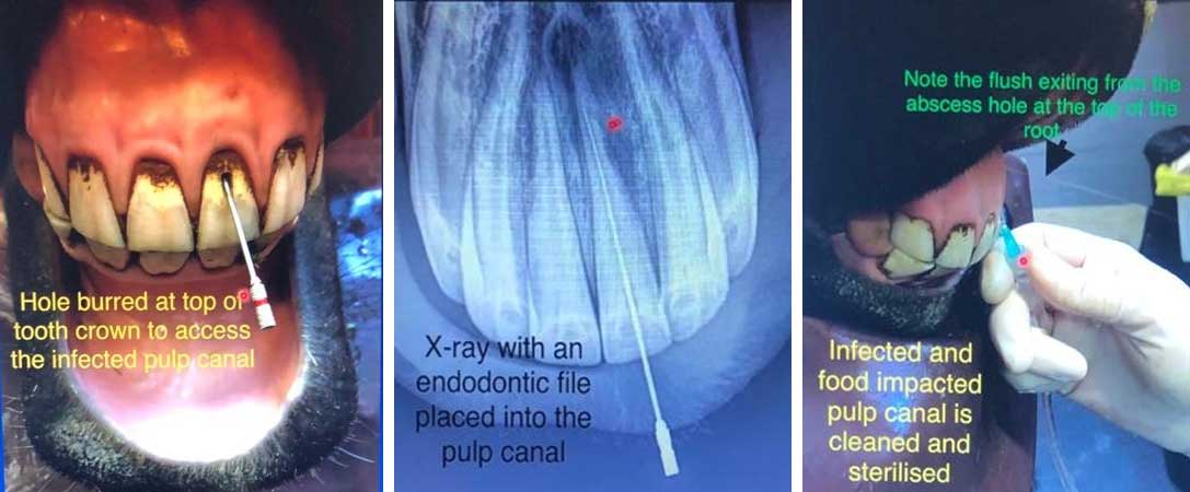 Root canal on incisors are fairly easy as there is only one pulp horn. From the three images above, the first shows a hole having been burred at the top of the tooth crown to access the infected pulp canal. The second photo is an x-ray showing the damage up at the root and the third photo is them flushing the infected pulp canal – if you look closely, the flush is exiting at the top through the abscess hole. Once the cleaning process is complete, a temporary filling is put in place where this continues to disinfect the canal for a further six weeks then a permanent filling will be put in.
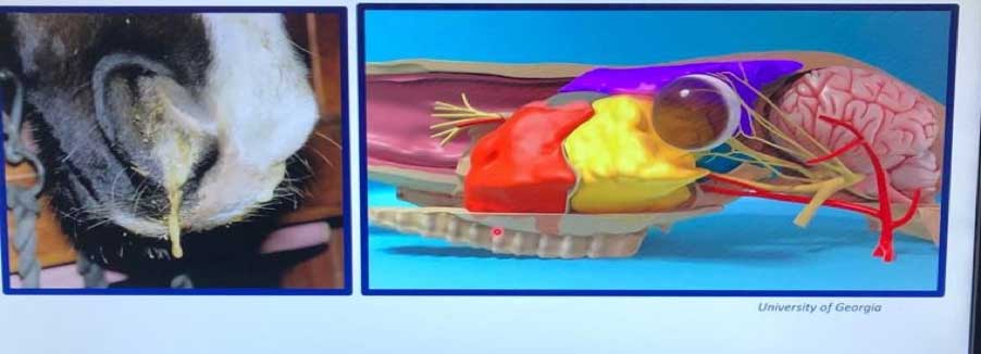
Sinusitis – a consequence of getting an infection in a cheek tooth is sinusitis. Sinuses are basically air-filled sacks that fill up the skull and the roots of the cheek teeth communicate with them. The first two cheek teeth go to the nasal cavity, but the third cheek tooth onwards communicate with the sinuses. If the pulp horns are exposed and not caught in time, the infection will bubble up into the sinuses. If you see discharge from the nose caused by dental infection, the horse will smell! To treat, you will need to remove the source (tooth) and the horse will need to have the sinus flushed.
 Infundibula Fractures are fairly common due to diastemas. Bacteria will feed off the tooth and will split the tooth in the middle. This is extremely uncomfortable for the horse and the split can also cause ulcers.
EORTH – Equine Odontoclastic Tooth Resorption & Hypercementosis. This is the reabsorption of the incisors. This can be really painful for the horse and is/can be found in the older horse. The roots get degraded and they can also lay down extra cement. You would do a carrot test. This is where the horse might not be able to bite into/snap the carrot. The teeth can also be wobbly and the only way to treat would be keep them clean or remove.
Two advanced techniques:
- MTE – Minimally Invasive Transbuccal Extraction. Come in through the side of the cheek/gum. Tap into the tooth and pull out
- Apicectomy – “retrograde endodontic and replantation procedure” take tooth out, cut top apex of tooth off and pull out the pulp horns, then put the tooth back in. Currently there is about a 30% success rate with this modern technique.
CASE STUDY – JEN’S HORSE, HARVEY
Saw Harvey a year ago and he was having a bit of trouble and was biting his rugs which can be a sign of pain/uncomfortable and he was also quidding. Upon investigations, they found he had diastema particularly in the mandibula teeth one of which was a bit offset. They tried a couple of times that year to clean and straighten his teeth, however, the problems were only alleviated for a short period of time before poor Harvey was then having problems again, so Alice took the tooth out.
On Harvey’s 300’s there was a tooth jutting out so in front and behind Harvey had two diastemas’, one was severe periodontal disease, and one was mild periodontal disease.
On his 400’s, Harvey had a tooth jetting out even further and Harvey had two severe diastemas. This offending tooth was removed. After the tooth was removed, a plug was put in to stop food collecting in the gap. This is then removed on a weekly basis for cleaning and trimming where the site heals. When it reaches 30% healed, the plug will be left out. Below you can see the healing process of Harvey.
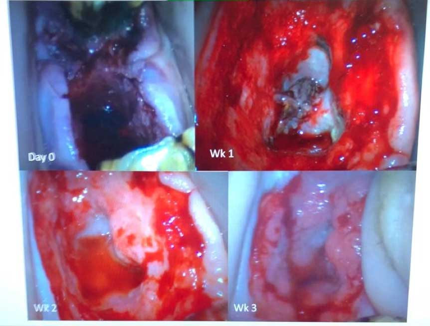
Harvey is now recovered and doing well which is great as this only happened on 28th January!!
Report by Charlotte Tarrant BHSI